Cbct In Endodontics Ppt
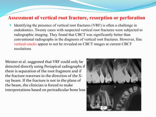
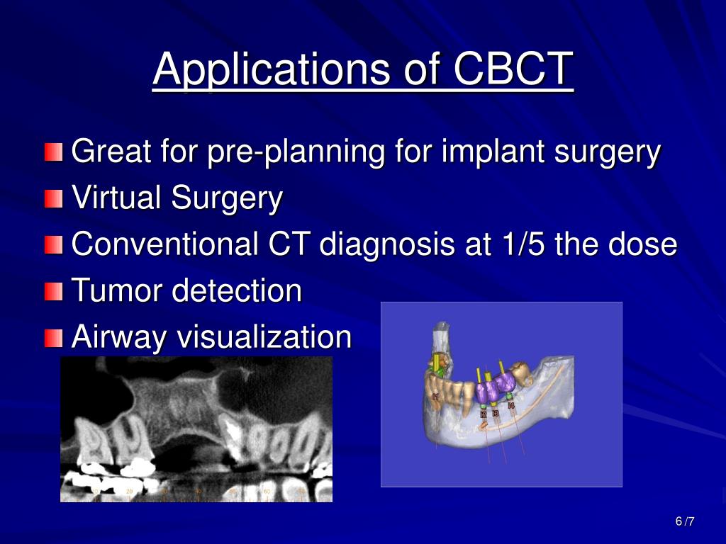
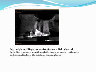
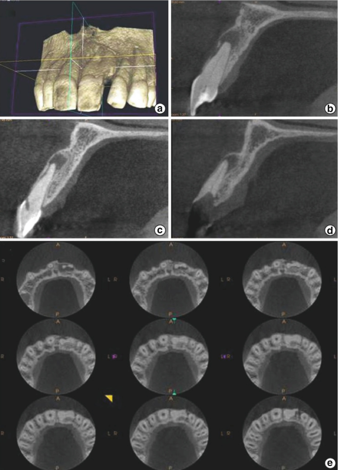
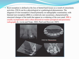
Cbct in endodontics ppt. Only small FOV CBCT scans are recommended for the diagnosis and management of endodontic problems. This clinical article aims to explain the benefits and limitations of CBCT and digital imaging. Cone beam computed tomography CBCT is a contemporary three- dimensional diagnostic imaging system designed specifically for use on maxillofacial skeleton It has its origins in.
To maximize the use of this imaging technology endodontists must have an in-depth knowledge and active role in each phase of CBCT imaging workflow which includes four. Gives the dentist confidence. 2 After a single two-dimensional projection is acquired by the detector the x-ray source and.
There are four primary areas in todays endodontic practice that can implement CBVI on a regular if not daily basis. Interpretation of cone beam computed tomography cbct f a 3-d conepyramid shaped divergent x-ray beam is directed through the patient onto a detector during exposure the x-ray generator. Enhances reputation patient engagement education and treatment understanding and acceptance.
Cbct scanner market insights forecast to 2025 - CBCT Scanner market is valued at million US in 2017 and will reach million US by the end of 2025 growing at a CAGR of during 2018-2025. HOW DOES CBCT WORK. The study of the tooth with CBCT is essential in cases where the anatomy of the tooth shows abnormalities in the radiographic study.
CBCT provides two features for orthodontic practice. Endodontic Practice US subscribers can answer. Cone beam computed tomography CBCT generates 3D volumetric images and is commonly used in dentistry 8.
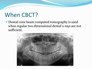
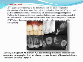
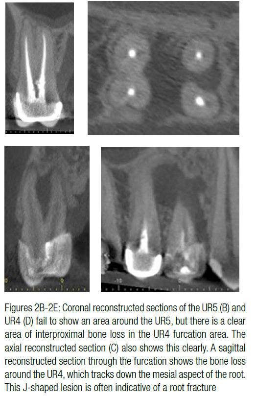
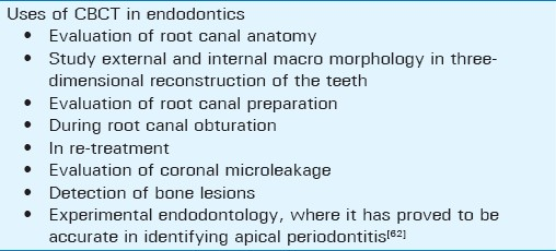
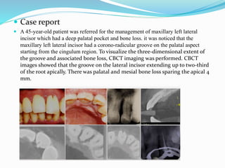
Advantages of CBCT in Endodontics Perhaps the most important advantage of CBCT in endodontics is that it demonstrates anatomic features in three dimensions that intraoral and. In such a case a CBCT scan is requested. A small FOV scan reduces the volume of exposed tissue and.
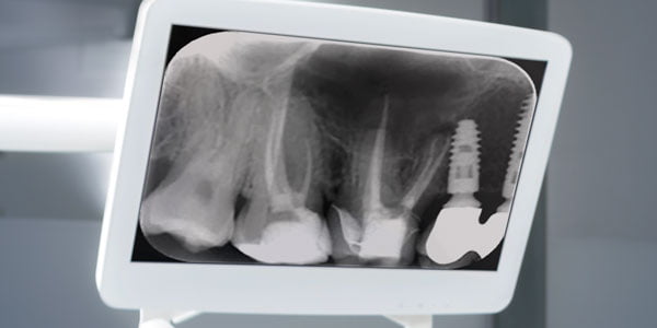
Specifically but not limited to these four areas is the. Estrela et al 2008 analyzed the accuracy of 1508 CBCT images periapical and panoramic radiographs for AP.
To maximize the use of this imaging technology endodontists must have an in-depth knowledge and active role in each phase of CBCT imaging workflow which includes four.
2 After a single two-dimensional projection is acquired by the detector the x-ray source and. 1 linear lateral and postero-anterior cephalometric images or curved planar projections panoramic images can be. They suggested that CBCT was an accurate diagnostic method to detect AP. Interpretation of cone beam computed tomography cbct f a 3-d conepyramid shaped divergent x-ray beam is directed through the patient onto a detector during exposure the x-ray generator. Gives the dentist confidence. 1 A 3D cone beam is directed through a central object onto a detector. There are four primary areas in todays endodontic practice that can implement CBVI on a regular if not daily basis. HOW DOES CBCT WORK. Endodontic Practice US subscribers can answer.
Enhances reputation patient engagement education and treatment understanding and acceptance. 1 linear lateral and postero-anterior cephalometric images or curved planar projections panoramic images can be. Advantages of CBCT in Endodontics Perhaps the most important advantage of CBCT in endodontics is that it demonstrates anatomic features in three dimensions that intraoral and. They suggested that CBCT was an accurate diagnostic method to detect AP. Cone beam computed tomography CBCT generates 3D volumetric images and is commonly used in dentistry 8. CBCT provides two features for orthodontic practice. Specifically but not limited to these four areas is the.


















Post a Comment for "Cbct In Endodontics Ppt"